Foot and Ankle

Feet are the foundation of our bodies and play an important part of a happy, healthy lifestyle. The foot is a complex structure that consists of 26 bones, 33 joints, and over 100 muscles, tendons, and ligaments. Its unique design allows the foot to handle hundreds of tons of force every day. The average adult takes 4,000 to 6,000 steps per day. That’s enough steps to walk around the earth four times during your life. When you consider the weight and stress we place on our feet each day, it’s easy to see how approximately 80% of people will experience a foot related problem at some point during their life.

ANATOMY

Each foot is made up of 26 bones, 33 joints and more than 100 muscles, tendons and ligaments, all of which work together to provide support, balance, and mobility. The foot can be put into three categories: the forefoot (metatarsals and phalanges), midfoot (cuboid, navicular and 3 cuneiforms), and hind foot (talus and calcaneus). Here’s a look at the main structures of the feet.

BONES

Approximately 25% of the bones in the body are found in our feet. These bones can be grouped into three parts:

  • Tarsal Bones: a set of seven irregularly shaped bones of the midfoot that form the foot’s arch. The tarsal bones include the calcaneus, talus, cuboid, navicular, and the medial, middle, and lateral cuneiforms.
    • Calcaneus: the heel bone and the largest bone of the foot.
    • Talus: also called the ankle bone, sits above the heel bone (calcaneus) and makes up the lower part of the ankle joint by connecting the tibia and fibula with the foot.
    • Cuboid: a cube-shaped bone that connects the foot to the ankle and helps provide stability to the foot.
    • Navicular: a boat-shaped bone that helps connect the talus (ankle bone) to the cuneiform bones.
    • 3 Cuneiform Bones: includes the medial, intermediate, and lateral cuneiform bones located between the navicular and first three metatarsals.

  • Metatarsals: five long bones of the forefoot that connect the tarsal bones with the toe bones (phalanges). Metatarsals are recognized by numbers, labeled one through five, starting with the big toe.
  • Phalanges: the 14 bones that make up the toes. Each toe consists of three phalanges, which consist of a proximal base, a middle shaft, and a distal head. However, the big toe (hallux) only has two phalanges, the distal and proximal.
    • Sesamoids: two small, pea-shaped bones embedded within a tendon beneath the big toe and provide a smooth surface for tendons to slide over.
    JOINTS

    Joints in the feet are formed wherever two or more of these bones meet. A layer of cartilage covers the articulating surfaces where two bones meet to form a joint. This allows bones to glide smoothly against one another as they move. The main Joints of the foot include:

    • Talocrural joint, or ankle joint, is formed between the tibia and fibula (bones of the lower leg) and the talus bone of the foot. It acts as a hinge joint, and allows for dorsiflexion (upward movement) and plantar flexion (downward movement).
    • Subtalar joint: also known as the agility joint, this joint formed between the talus and the calcaneus.
    • Transverse tarsal joint: is a combination of the talocalcaneonavicular joint and the calcaneocuboid joint.
    • Talocalcaneonavicular joint: is formed between the talus, calcaneus, and navicular bones.
    • Calcaneocuboid joint: is formed between the front of the calcaneus and the posterior surface of the cuboid bone.
    • Cuneonavicular joint: is formed between the navicular bone and the three cuneiform bones.
    • Cuboideonavicular joint: is formed between the cuboid and navicular bones.
    • Tarsometatarsal joints: are formed between the tarsal bones and the bases of the metatarsal bones.
    • Intermetatarsal joints: involve the bases of the metatarsal bones.
    • Interphalangeal joints: These joints connect the phalanges. They’re synovial joints strengthened by collateral and plantar ligaments, and they let you flex and extend your toes.
    • Metatarsophalangeal joint (MCP): the joint at the base of the toe.
    • Proximal interphalangeal joint (PIP): the joint in the middle of the toe.
    • Distal phalangeal joint (DP): the joint closest to the tip of the toe.
    MUSCLES

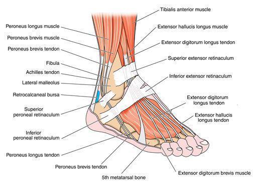
    Twenty muscles give the foot its shape, support, and ability to move. The main muscles of the foot are:

    • Posterior tibilias supports the foot’s arch.
    • Anterior tibilias allows the foot to move upward.
    • Peroneal tibilias controls the movement on the outside of the ankle.
    • Extensors raise the toes, making it possible to take a step.
    • Flexors stabilize the toes.
    TENDONS AND LIGAMENTS

    Tendons attach muscles to the bones, while ligaments attach bones to other bones. Tendons and ligaments work together to provide movement, stabilize the joints, and maintain anatomical structures in our feet. The main tendons of the foot include:

    • Achilles tendon: attaches the calf muscle to the heel bone. The Achilles tendon makes it possible to run, jump, climb stairs and stand on your toes.
    • Posterior tibial tendon: attaches one of the smaller muscles of the calf to the underside of the foot. This tendon helps support the arch and allow us to turn the foot inward.
    • Anterior tibial tendon: allows us to raise the foot.
    • Lateral malleolus: two tendons that run behind the out bump of the ankle and help us turn the foot out.
    The main ligaments of the foot are
    • Plantar fascia: the longest ligament of the foot. The ligament, which runs along the sole of the foot, from the heel to the toes, forms the arch. By stretching and contracting, the plantar fascia helps us balance and gives the foot strength for walking.
    • Plantar calcaneonavicular ligament– a ligament of the sole of the foot that connects the calcaneus and navicular and supports the head of the talus.
    • Calcaneocuboid ligament– the ligament that connects the calcaneus and the tarsal bones and helps the plantar fascia support the arch of the foot.
    FOOT AND ANKLE CONDITIONS & INJURIES